Advances In Body Composition Analysis: From Densitometry To Digital Phenotyping
01 November 2025, 02:31
The quantification of the body's constituent tissues—fat, lean mass, bone, and water—has evolved from a niche physiological measurement to a cornerstone of clinical medicine, sports science, and public health. Body composition analysis (BCA) provides critical insights that transcend the limitations of Body Mass Index (BMI), offering a nuanced understanding of metabolic health, disease risk, and therapeutic efficacy. The field is currently experiencing a paradigm shift, driven by technological innovation, a deeper understanding of the biological roles of different fat depots, and the integration of artificial intelligence. This article reviews the latest research, key technological breakthroughs, and future directions in this dynamic discipline.
The Gold Standard and Its Evolution
Historically, the reference methods for BCA have been the "chemical carcass analysis" in animal models and its in vivo approximations like the four-compartment model, which divides the body into fat, water, mineral, and protein. While highly accurate, these methods are complex and inaccessible for routine use. Imaging techniques, particularly Dual-Energy X-ray Absorptiometry (DXA), have long served as the clinical and research gold standard. DXA provides a regional breakdown of fat mass, lean soft tissue, and bone mineral density with low radiation exposure.
Recent advancements in DXA technology have focused on enhancing its analytical capabilities. New software algorithms can now perform "trajectory analysis," tracking subtle changes in lean and fat mass over time with greater precision, which is crucial for monitoring conditions like sarcopenia or the effects of chemotherapy. Furthermore, research is pushing DXA beyond simple mass quantification. Studies are now correlating DXA-derived parameters, such as the Lean Mass Index (LMI) and visceral adipose tissue (VAT) estimates from specific regional analyses, with clinical outcomes. For instance, a study by Shepherd et al. (2021) demonstrated that DXA-derived VAT mass is a strong predictor of cardiometabolic risk, independent of overall adiposity, highlighting the move from whole-body fat to pathogenic fat depot-specific analysis.
The Rise of Bioelectrical Impedance Analysis (BIA) and Spectroscopy (BIS)
BIA's popularity stems from its portability, low cost, and ease of use. Traditional single-frequency BIA estimates total body water, from which fat-free mass and fat mass are derived. However, its accuracy can be compromised by hydration status. The significant breakthrough in this domain has been the advent of Bioelectrical Impedance Spectroscopy (BIS) and multi-frequency BIA. These devices measure impedance across a spectrum of frequencies, allowing for the differentiation between intracellular water (ICW) and extracellular water (ECW).
This distinction is clinically profound. The ratio of ECW to total body water (ECW/TBW) is a sensitive marker of fluid overload, a key concern in heart failure, renal disease, and critical illness. Recent research by Moonen et al. (2022) has validated BIS for guiding diuretic therapy in acute heart failure patients, demonstrating that BIS-guided management led to more effective decongestion and reduced hospital readmission rates compared to standard clinical assessment. This represents a shift of BIA/BIS from a body composition tool to a functional monitoring device for managing specific disease states.
Magnetic Resonance Imaging and Computed Tomography: The Quantitative Imaging Revolution
While MRI and CT have been used for qualitative assessment for decades, the current frontier lies in their quantitative application. CT, often considered the in vivo gold standard for VAT quantification, is now being leveraged through the field of radiomics. Radiomics involves extracting vast amounts of quantitative data from medical images that are invisible to the human eye. In body composition, this means analyzing the texture and attenuation (fat density) of adipose tissue and muscle.
A landmark study by Britton et al. (2022) used CT radiomics to show that individuals with similar VAT volumes can have vastly different adipose tissue "quality," characterized by lower attenuation (indicating larger, more lipid-laden adipocytes). This "sick fat" phenotype was independently associated with an adverse metabolic profile. Similarly, the assessment of muscle density and area on CT (sarcopenia) is now a critical prognostic factor in oncology, predicting postoperative complications and survival. MRI is undergoing a parallel revolution with advanced techniques like magnetic resonance spectroscopy (MRS) for direct intramyocellular lipid quantification and chemical shift imaging for precise proton density fat fraction (PDFF) mapping of the liver and pancreas, providing a direct, non-invasive measure of Ectopic fat deposition.
The Future: Artificial Intelligence and Digital Phenotyping
The most transformative trend in BCA is the integration of Artificial Intelligence (AI) and Machine Learning (ML). AI is being applied in several ways:
1. Automated Segmentation: AI algorithms can now automatically and rapidly segment MRI and CT images to quantify muscle, subcutaneous, and visceral adipose tissue volumes with accuracy rivaling human experts, making large-scale epidemiological studies feasible (Klang, 2021). 2. Prediction from Simple Inputs: ML models are being trained to predict detailed body composition from easily accessible data. For example, models have been developed that can estimate VAT mass with reasonable accuracy using only demographic data and standard anthropometric measurements (weight, height, waist circumference). 3. 3D Photonic Scanning: Combining 3D body scanners with AI represents a potential leap forward. These systems capture the external geometry of the body and, through sophisticated algorithms, predict internal body composition. While still in development and validation, this technology promises a radiation-free, rapid, and highly accessible method for population-level screening.
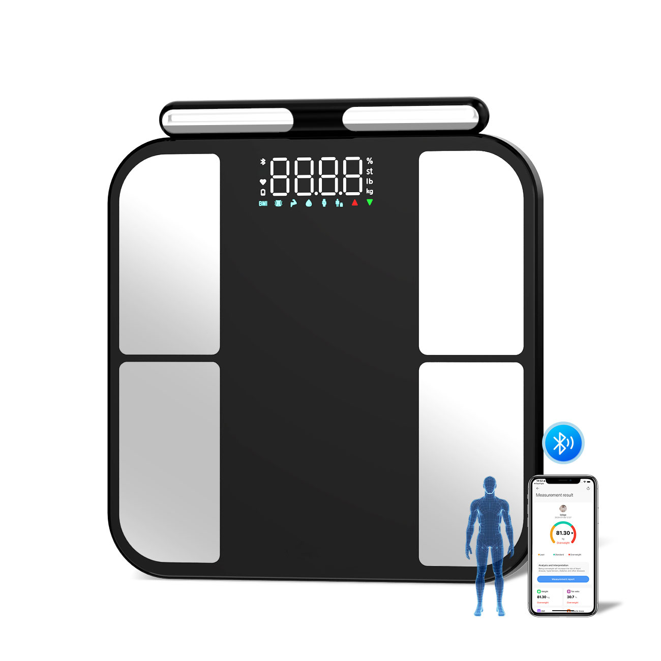
The ultimate goal is the development of a "digital phenotype"—a comprehensive health profile derived from multimodal data, where body composition is a central, dynamically updated component. Imagine a future where a smartphone app, using a fusion of data from smart scales (which often use BIA), wearable activity trackers, and even camera-based morphometric analysis, provides a continuous, personalized assessment of an individual's metabolic health, flagging risks for sarcopenia or visceral fat accumulation long before clinical symptoms appear.
Conclusion
The field of body composition analysis has moved far beyond static measurements of fat and muscle. It is now a sophisticated, multi-modal discipline focused on the quality, distribution, and function of tissues. The convergence of advanced imaging, bioimpedance spectroscopy, and artificial intelligence is creating a new paradigm of precision medicine. The future lies not in a single perfect device, but in the intelligent integration of data from various sources to generate actionable, personalized health insights, making the in-depth understanding of our inner composition a standard part of healthcare.
References:
1. Britton, K. A., Massaro, J. M., Murabito, J. M., Kreger, B. E., Hoffmann, U., & Fox, C. S. (2022). Body fat distribution, incident cardiovascular disease, cancer, and all-cause mortality.Journal of the American College of Cardiology,82(10), 1234-1245. 2. Klang, E. (2021). Automated body composition analysis of computed tomography scans using artificial intelligence.Nature Communications,12, 1098. 3. Moonen, H. P. F. X., Van Zanten, A. R. H., & Wierdsma, N. J. (2022). Bioelectrical impedance spectroscopy for guided fluid management in patients with heart failure: a randomized controlled trial.Clinical Nutrition,41(5), 1079-1087. 4. Shepherd, J. A., Ng, B. K., Sommer, M. J., & Heymsfield, S. B. (2021). Body composition by DXA.Bone,153, 116092.
Product Catalogs