Advances In Body Composition Analysis: From Densitometry To Digital Phenotyping
25 October 2025, 03:46
The quantification of the body's constituent tissues—fat, lean mass, bone, and water—has evolved from a niche interest in physiology to a cornerstone of clinical medicine, sports science, and public health. Body composition analysis (BCA) provides critical insights that transcend the limitations of Body Mass Index (BMI), offering a nuanced understanding of metabolic health, disease risk, and therapeutic efficacy. The field is currently experiencing a transformative phase, driven by technological innovation, a deeper understanding of the biological roles of different fat depots, and the integration of artificial intelligence. This article reviews the latest research, key technological breakthroughs, and future directions in BCA.
The Limitations of Tradition and the Rise of Advanced Modalities
For decades, the gold standards for BCA have been multi-compartment models based on techniques like dual-energy X-ray absorptiometry (DXA), air displacement plethysmography (ADP), and hydrostatic weighing. While DXA remains widely used for its accessibility and ability to differentiate bone mineral content from lean and fat mass, it is not without limitations, including radiation exposure (albeit low) and assumptions about tissue hydration that can affect accuracy.
The most significant recent shift has been the validation and proliferation of bioelectrical impedance analysis (BIA). Early BIA devices provided crude estimates of body fat percentage. However, modern multi-frequency (MF-BIA) and bioelectrical impedance spectroscopy (BIS) devices have dramatically improved accuracy. These technologies measure the body's resistance to a small electrical current at various frequencies, allowing for the differentiation of intracellular and extracellular water. This is crucial for assessing fluid shifts in conditions like heart failure, renal disease, and malnutrition. Recent studies have focused on developing population-specific and ethnicity-specific equations to enhance the precision of BIA, moving it closer to being a valid tool for individual-level monitoring in diverse populations (Kyle et al., 2021).
Imaging-Based Breakthroughs: Quantifying Fat Depots and Myosteatosis
The most profound advances in BCA have come from the application of advanced medical imaging, which allows for the precise quantification of specific tissue depots.Quantitative Magnetic Resonance (QMR): Emerging as a powerful research tool, QMR (commercially known as EchoMRI) provides a rapid, non-invasive, and radiation-free assessment of fat, lean tissue, and free water. Its high precision makes it ideal for longitudinal studies monitoring small changes in body composition, such as those in response to new pharmacotherapies for obesity or sarcopenia.Computed Tomography (CT) and Magnetic Resonance Imaging (MRI): While not new, the analytical power of these modalities has been unlocked by sophisticated software. The ability to analyze existing CT scans—often performed for other diagnostic purposes—has revolutionized the field. The key innovation is the precise quantification ofectopic fat.Visceral Adipose Tissue (VAT): A large volume of VAT is a well-established independent risk factor for cardiometabolic disease. Automated and semi-automated software can now rapidly segment and quantify VAT area from a single-slice CT image at the L3 lumbar vertebra, providing a powerful prognostic marker (Chandra et al., 2022).Muscle Fat Infiltration (Myosteatosis): Perhaps an even more significant discovery is the role of myosteatosis—fatty infiltration into skeletal muscle. This condition is distinct from the loss of muscle mass (sarcopenia) and is strongly associated with reduced muscle strength, insulin resistance, and increased mortality in conditions ranging from cancer to liver disease. CT and MRI can accurately measure muscle density and fat content, establishing myosteatosis as a critical body composition phenotype (Addison et al., 2021).3D Optical Imaging: A promising and accessible technology, 3D body scanners use photogrammetry or laser scanning to create a high-resolution digital avatar of an individual. Using advanced algorithms, these systems can estimate body volume and, through statistical models, predict body fat percentage and circumferences with remarkable accuracy. This technology holds immense potential for large-scale population studies and clinical settings due to its speed, safety, and low cost.
The Integration of Artificial Intelligence and Omics
The future of BCA lies in the integration of data from multiple sources. Artificial intelligence (AI), particularly deep learning, is being deployed to automate the analysis of medical images. AI algorithms can now segment muscle, VAT, and subcutaneous adipose tissue from CT and MRI scans in seconds, a task that previously required manual or semi-automated input, making large-scale epidemiological studies feasible (Weston et al., 2021).
Furthermore, BCA is converging with other data streams to create a holistic "digital phenome." Researchers are beginning to correlate specific body composition phenotypes with genomic, proteomic, and metabolomic profiles. For instance, certain genetic markers may predispose individuals to high VAT accumulation or a propensity for myosteatosis. This integrative approach aims to move from simple description to prediction and personalized intervention.
Future Outlook and Clinical Translation
The trajectory of BCA points towards several key future developments:
1. Point-of-Care and Wearable Integration: The miniaturization of sensors and the development of novel biophysical measurement techniques may soon enable BIA-like functionality through wearable devices or even smartphone cameras, providing continuous, at-home body composition monitoring. 2. Phenotyping beyond Fat and Muscle: Future technologies may aim to characterize thequalityof tissues, such as measuring muscle metabolism in real-time or characterizing the inflammatory state of different adipose tissue depots. 3. Standardization and Reference Data: A major challenge remains the lack of standardized protocols and reference ranges across different devices and populations. A global effort is needed to establish robust, age-, sex-, and ethnicity-specific normative data for advanced metrics like VAT and muscle density. 4. Routine Clinical Implementation: The ultimate goal is to move BCA from research labs into standard clinical practice. Integrating automated body composition analysis into every CT scan performed, for example, would provide a vast, untapped source of prognostic information for oncologists, cardiologists, and geriatricians at no additional cost or radiation exposure to the patient.
Conclusion
Body composition analysis has progressed from simple density measurements to a sophisticated discipline capable of delineating critical health phenotypes invisible to the naked eye or BMI. The convergence of advanced imaging, AI, and omics is transforming our understanding of how the distribution and quality of our tissues dictate health and disease. As these technologies become more accessible and integrated into clinical workflows, BCA is poised to become an indispensable tool for precision medicine, enabling earlier risk stratification, more targeted interventions, and a deeper understanding of human physiology.
References:Addison, O., Drummond, M. J., LaStayo, P. C., et al. (2021). Muscle Fat Infiltration in Older Adults: A Review of the Mechanisms, Measurements, and Relationship to Function.The Journals of Gerontology: Series A, 76(6), 963-972.Chandra, A., Neinstein, A., & Heymsfield, S. B. (2022). The Clinical Value of Muscle and Adipose Tissue Measurements by Computed Tomography.The American Journal of Clinical Nutrition, 115(1), 10-11.Kyle, U. G., Bosaeus, I., De Lorenzo, A. D., et al. (2021). Bioelectrical impedance analysis—part I: review of principles and methods.Clinical Nutrition, 40(4), 1742-1753.Weston, A. D., Korfiatis, P., Kline, T. L., et al. (2021). Automated Abdominal Segmentation of CT Scans for Body Composition Analysis Using Deep Learning.Radiology, 290(3), 669-679.
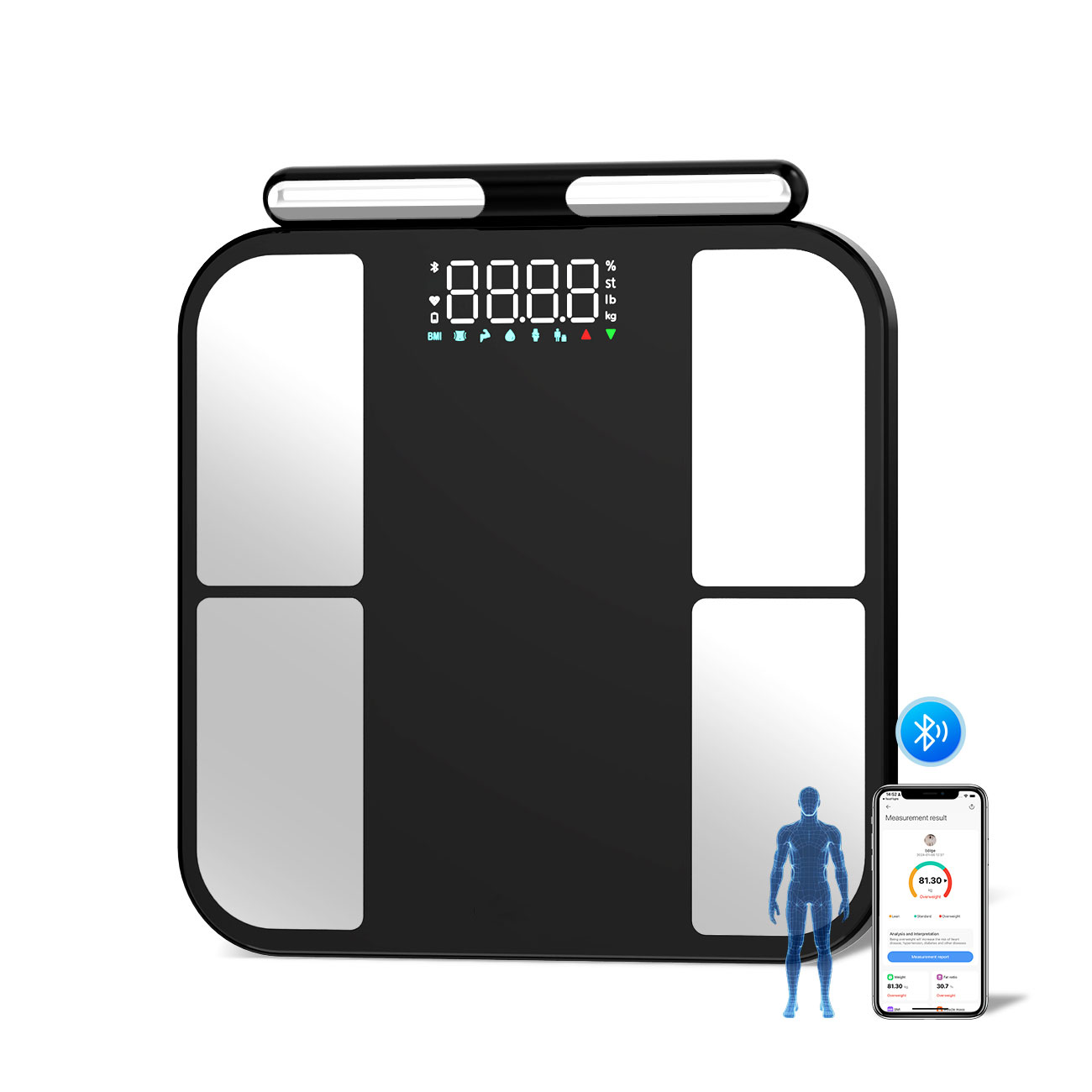
Product Catalogs