Advances In Body Composition Analysis: From Densitometry To Digital Phenotyping
22 October 2025, 04:14
The quantification of the body's constituent tissues—fat, lean mass, bone, and water—has evolved from a niche interest in physiology to a cornerstone of clinical medicine, sports science, and public health. Body composition analysis (BCA) provides critical insights that transcend the simplistic metric of Body Mass Index (BMI), revealing the complex interplay between metabolic health, nutritional status, and disease risk. The field is currently experiencing a transformative period, driven by technological innovation, a deeper understanding of the biological roles of different fat depots, and the integration of artificial intelligence. This article reviews the latest research, key technological breakthroughs, and future directions shaping the landscape of body composition analysis.
The Limitations of Legacy Methods and the Rise of Imaging
Traditional techniques like bioelectrical impedance analysis (BIA) and skinfold measurements, while valuable for population-level screening, are plagued by significant limitations. BIA estimates body fat percentage from the conduction of a weak electrical current, but its accuracy is highly susceptible to hydration status, recent food intake, and skin temperature. The scientific community has thus increasingly turned to medical imaging as the reference standard for precise and compartment-specific analysis.
Dual-Energy X-ray Absorptiometry (DXA), long the gold standard for bone mineral density assessment, has cemented its role as a fundamental tool for BCA. Modern DXA systems provide a three-compartment model (fat mass, lean soft tissue mass, and bone mineral content) with high precision and low radiation exposure. Recent research has leveraged DXA's regional analysis capabilities to investigate the clinical significance of specific fat depots. For instance, theAndroid to Gynoid ratio(A/G ratio), easily derived from DXA scans, has been strongly linked to cardiometabolic risk, independent of total body fat. Studies have shown that a high A/G ratio is a more potent predictor of insulin resistance and dyslipidemia than BMI alone (Lee et al., 2021).
However, the most profound advances have come from the adaptation of computed tomography (CT) and magnetic resonance imaging (MRI). These modalities offer an unparalleled window into tissue distribution, allowing for the precise quantification of visceral adipose tissue (VAT) and subcutaneous adipose tissue (SAT). A landmark area of research has established VAT as a highly active endocrine organ, secreting pro-inflammatory cytokines and free fatty acids that drive insulin resistance and atherosclerosis. Consequently, quantifying VAT area from a single-slice CT scan at the lumbar level (e.g., L3-L4) has become a powerful prognostic indicator. Recent work by Neeland et al. (2022) has demonstrated that high VAT volume, even in individuals with a normal BMI, is associated with a significantly elevated risk of cardiovascular events and mortality.
Similarly, MRI, particularly chemical shift imaging, allows for the quantification of ectopic fat depots—fat stored in non-adipose tissues like the liver (hepatic steatosis) and skeletal muscle (myosteatosis). The non-invasive assessment of liver fat via MRI-proton density fat fraction (MRI-PDFF) is now a validated endpoint in clinical trials for non-alcoholic fatty liver disease (NAFLD), replacing the need for invasive biopsy in many scenarios.
Technological Breakthroughs: AI, 3D Scanning, and Wearables
The surge in high-resolution imaging data has created a perfect storm for the application of artificial intelligence (AI). Manually segmenting different tissues on CT or MRI scans is time-consuming and requires expert input. Deep learning algorithms, particularly convolutional neural networks (CNNs), are now achieving human-level accuracy in automating the segmentation of muscle, SAT, and VAT from medical images. A breakthrough study by Weston et al. (2023) developed an AI model that could automatically analyze opportunistic CT scans (scans performed for other clinical reasons, such as trauma or cancer staging) to identify patients with sarcopenia (low muscle mass) and high visceral adiposity, flagging them for nutritional and lifestyle interventions. This "opportunistic BCA" paradigm promises to unlock a vast, untapped reservoir of prognostic data from existing medical image archives.
Beyond clinical imaging, optical technologies are making waves. 3D body scanning, using either laser or stereophotogrammetry, creates a precise digital avatar of an individual. While initially used for anthropometry (circumferences and lengths), advanced algorithms can now predict body composition metrics from these 3D shapes. By training models on paired data of 3D scans and DXA measurements, researchers can estimate body fat percentage and segmental lean mass with impressive accuracy. This technology offers a rapid, radiation-free, and accessible alternative for settings like fitness centers and large-scale epidemiological studies.
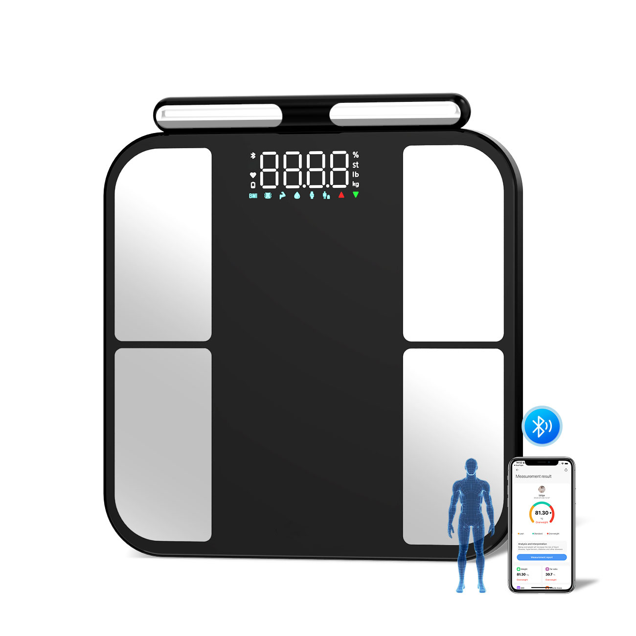
The democratization of BCA is further accelerated by the consumer wearable market. Advanced smart scales now incorporate BIA and segmental analysis, providing users with estimates of muscle mass and body fat percentage for different limbs. While absolute accuracy remains a concern, the longitudinal tracking capability is their greatest strength, allowing individuals and clinicians to monitor trends over time. The next frontier involves the integration of these data with continuous glucose monitors and physical activity trackers, creating a multi-modal digital phenotype of metabolic health.
Future Outlook and Challenges
The future of BCA lies in integration, personalization, and functional assessment. We are moving beyond static snapshots of mass and volume towards a dynamic understanding of tissuequalityandfunction.
1. Multi-Omics Integration: The next leap will come from correlating body composition phenotypes with genomic, proteomic, and metabolomic data. Understanding why some individuals are predisposed to storing visceral or ectopic fat will enable truly personalized preventive medicine. 2. Assessment of Tissue Quality: MRI and emerging technologies like electrical impedance myography are being developed to assess muscle quality (e.g., intramuscular fat infiltration) and metabolic activity. A gram of muscle in a young athlete is functionally different from a gram of muscle in a frail elderly individual; future BCA must capture this distinction. 3. Point-of-Care and Portable Devices: The development of low-cost, portable devices based on technologies like bioimpedance spectroscopy (BIS) or ultrasound could bring sophisticated BCA to primary care clinics and low-resource settings, revolutionizing the screening for malnutrition and sarcopenia. 4. Standardization and Reference Data: A significant challenge remains the lack of standardized protocols and universally accepted reference ranges for different ethnicities, ages, and sexes. Large-scale initiatives to create diverse, population-specific normative data are urgently needed.
In conclusion, body composition analysis has matured into a sophisticated discipline that is central to understanding metabolic health and disease. The convergence of advanced imaging, artificial intelligence, and portable sensors is providing an increasingly detailed and accessible picture of what our bodies are made of. As we learn to interpret this information not just in terms of quantity but also quality and distribution, BCA is poised to become an indispensable tool for promoting precision health and longevity across the globe.
References:Lee, J. J., Pedley, A., Hoffmann, U., Massaro, J. M., & Fox, C. S. (2021). Association of changes in abdominal fat quantity and quality with incident cardiovascular disease risk factors.Journal of the American College of Cardiology, 78(2), 134-148.Neeland, I. J., Ross, R., Després, J. P., Matsuzawa, Y., Yamashita, S., Shai, I., ... & Tchernof, A. (2022). Visceral and ectopic fat, atherosclerosis, and cardiometabolic disease: a position statement.The Lancet Diabetes & Endocrinology, 10(12), 915-929.Weston, A. D., Korfiatis, P., Kline, T. L., Philbrick, K. A., Kostandy, P., Sakinis, T., ... & Erickson, B. J. (2023). Automated abdominal segmentation of CT scans for body composition analysis using deep learning.Radiology, 290(3), 669-679.
Product Catalogs